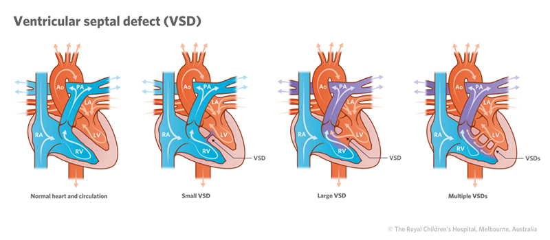
A ventricular septal defect (VSD) is a hole in the wall of the heart between the pumping chambers of the heart (ventricles). A VSD allows oxygen-rich “red” blood to pass from the left ventricle through the hole and mix with oxygen-poor “blue” blood in the right ventricle.
Mixing of the blood causes changes to oxygen levels in the body and the heart has to work harder to maintain the body’s requirements. It also causes an increase in blood flow to the lungs and may expose the lungs to high pressure.
A child may have several VSDs and they can vary in size. Most VSDs are small and spontaneously close within the first few years of life.
Signs and symptoms
The main signs and symptoms of VSD include:
- breathlessness (particularly when a baby feeds)
- sweating
- tiredness
- poor feeding
- slow weight gain.
Diagnosis
Tests to help diagnose VSD include:
- Electrocardiogram (ECG) – measures the electrical activity of the heart.
- Echocardiogram (Echo) – uses sounds waves to produce a moving picture of the heart.
- CT Scan – an X-ray with dye injected to highlight blood vessels.
- Cardiac catheterisation – A thin flexible tube (called a catheter) is inserted into a blood vessel in the groin and fed up into the heart to measure pressures and oxygen levels, and visualise heart structures using X-ray equipment. The procedure is performed under a general anaesthetic. Learn more about cardiac catheterisation.
These tests are usually carried out because a doctor has heard a heart murmur when examining a child with a stethoscope.
Treatment
The treatment for VSD may include medication and/or surgery.
Medications include:
- Diuretics – medication to help the kidneys remove excess fluid in the body.
- ACE inhibitors – medication to relax the blood vessels, making it easier for the heart to pump blood through the body.
- Nutritional supplements may also be required for babies who are not growing adequately.
Surgery options include:
- using a patch or stitch to close the hole
- inserting a device using a cardiac catheter. A special device is passed through the catheter into the VSD where it expands and plugs the hole.
Children with multiple VSDs may need a combination of the above.
In some cases, if the child is too young, a pulmonary artery band may be used to control the excessive blood flow through their lungs, giving them time to grow until they can have surgery to correct the VSD.