Introduction
Head injuries are a common paediatric ED presentation, accounting for 1 – 2% of all presentations to specialist children’s emergency services within Australia.1 Although most are minor, head injuries remain a significant cause of morbidity and mortality.
Children sustaining head injuries at the more severe end of the head injury spectrum are usually readily identifiable and this should prompt immediate (and concurrent) intervention, investigation and referral for definitive management.
Children with clinical features of head injury at the “milder”, and by far more prevalent end of the spectrum, present their own challenges and differentiating the child with the truly low risk head injury from those at risk of a clinically significant injury, such as an intracranial bleed or a depressed skull fracture, can be problematic. While a CT (Computed Tomography) scan is the investigation of choice to exclude such injuries in the acute setting, it is neither feasible nor ethical to scan every child presenting given concerns with radiation exposure, the potential need for sedation and/or transfer, and resource costs.
Several clinical decision rules (CDRs) have been derived to risk stratify children with isolated head injuries and thus to guide clinicians in imaging and discharge decisions. This guideline is informed by the highest performing CDRs in our setting and adopts the concept of the clinico-radiological rule to enable an evidence-based approach to the decision making around head injury management in Queensland.
Clinical decision rules
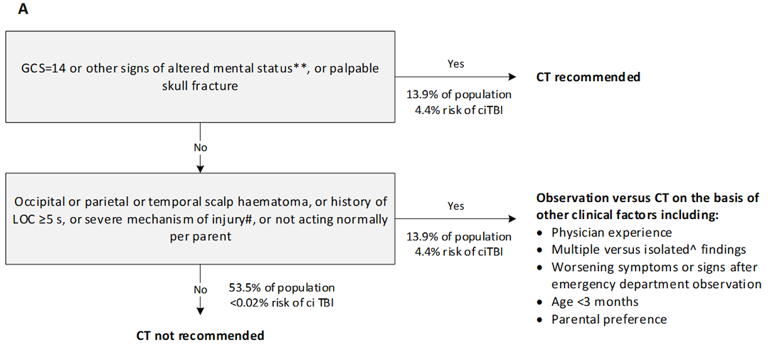
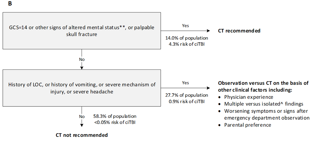
Clinical decision rules (CDRs) in paediatric head injury have been derived to guide imaging decisions. The most well known and highest quality CDRs are PECARN2 (US study, 33,785 children), CHALICE3 (UK, 22,772) and CATCH4 (Canada, 3688). PECARN and CHALICE CDRs are outlined below. PECARN is designed for children with a GCS >13, offers different rules for those less than two years and those greater than two years and is a clinico-radiological rule with the option to observe or image children at intermediate risk. CHALICE considers all children presenting with a head injury and CATCH was designed for a higher risk cohort. Both PECARN and CHALICE have a negative predictive value of 99.9%, meaning that children who are negative for the rule i.e. low risk, are estimated to have less than 0.1% risk of significant intracranial injury.
A recent Australia New Zealand observational multicentre study, APHIRST,5 examined the performance of all three of these CDRs in our context. All three rules performed well, PECARN had the highest sensitivity, and all three rules had a negative predictive value over 99% (PECARN – 100% (95%CIs 99.9-100; 99.8-100 for each age group); CHALICE 99.8% (95%CI 99.7-99.9). It is important to recognise though, that strict application of the rules would likely have resulted in a much higher imaging rate than currently exists in the 10 tertiary and large mixed hospitals that were part of the study. The APHIRST baseline imaging rate among all children was 10.5%; PECARN if strictly applied could result in an imaging rate as high as 46.6% (depending on whether intermediate risk children were observed or scanned), CHALICE 22% and CATCH 30%. Furthermore, in the APHIRST study, clinician gestalt in children with milder head injuries (GCS 13-15) was found to perform better than any rule (no missed injuries).6 The ten APHIRST sites are either tertiary children’s hospitals or large mixed centres with a strong paediatric focus, and clinician decisions are likely informed by factors including awareness of the CDRs, clinician experience, and the use of observation instead of immediate CT scan as a management option in some cases.
There is considerable overlap between the CDRs in clinical assessment variables (history, mechanism of injury, examination), albeit with some discrepancy over exact details e.g. height of fall, number of vomits and length of any loss of consciousness. These features and the CDRs have been used to inform the statewide guideline. Scope has also been given to allow for observation as an initial option in some children deemed at intermediate risk.
Assessment
Emergency care should always involve a rapid primary survey with evaluation of (and immediate management of concerns with) airway, breathing, circulation and disability (ABCD). This includes level of consciousness and Glasgow coma score (GCS).
The aim of the assessment is to:
- identify a child with a severe head injury at risk or showing signs of raised intracranial pressure (ICP) to enable immediate investigation, management and prompt referral
- differentiate children at low risk of a clinically significant head injury (who can be safely discharged without the need for a CT scan) from those who require further management (CT scan or observation).
- identify children with other concerns e.g. non-accidental injury (NAI), alternate diagnoses.
An integration of clinical assessment features into low, intermediate and high risk is included in head injury management (see table on risk stratification in Management section).
History and mechanism of injury
Important factors to elicit on history include:
- abnormal behaviour such as agitation or drowsiness
- loss of consciousness
- vomiting
- post traumatic seizure
- amnesia: retrograde and antegrade
- headache
- mechanism of injury – significant mechanisms of injury include:
- fall from a significant height
- motor vehicle accident (especially if high-speed, ejected from vehicle or others significantly injured in the same crash)
- pedestrian/cyclist impacted by a motor vehicle
- impact from high-speed projectile e.g. golf ball, ceiling fan
- any special circumstances to consider including injuries of potential NAI concern and alternate diagnoses (refer to Further Assessment Considerations below)
Some differences exist between CDRs in symptoms and signs included as important, and the degree e.g. height of fall, number of vomits, length of loss of consciousness.
Examination
Following the primary survey, a thorough head-to-toe examination (secondary survey) with specific attention paid to the maintenance of neutral positioning if cervical spine injury concerns (refer to Cervical spine injury guideline) should occur.
Particular features on examination to identify are:
- level of consciousness (LOC), including GCS or AVPU score. All CDRs consider that a child with a diminished or decreasing LOC is at significant risk of intracranial injury. If present, examination should be made for other signs of raised intracranial pressure (ICP)
- suspicion of an open or depressed skull fracture (including boggy haematomas, palpable depressions)
- signs of a basal skull fracture (raccoon eyes, haemotympanum, Battle’s sign, CSF leak via nose, ears)
- penetrating injury
- presence of focal neurological deficit
- other features suggestive of more extensive injury (e.g. other significant trauma, NAI)
- in infants and young children: size and location of a haematoma, swelling or laceration should be noted, as should a bulging fontanelle (see PECARN and CHALICE CDRs)
Signs of raised ICP
- deteriorating or diminished LOC
- abnormal posture (decorticate or decerebrate)
- abnormal pupillary responses, unilateral or bilateral dilatation
- abnormal oculocephalic reflexes (doll’s eye movement or dysconjugate upward gaze)
- abnormal breathing patterns (hyperventilation, Cheyne-Stokes, apnoea)
Cushing’s triad (hypertension + bradycardia + breathing abnormalities) is a late sign.
Careful initial and repeated clinical examination is required to identify signs of raised ICP.
Seek urgent paediatric critical care/neurosurgical advice for a child with signs of raised ICP or decreased level of consciousness (onsite or via Retrieval Services Queensland (RSQ).
Further assessment considerations
Consider special circumstances in the presentation, or the possibility that the clinical presentation is unrelated to the head injury.
| Further assessment considerations in children presenting with head injury |
|---|
| Non-accidental Injury (NAI) |
Concerns of NAI necessitate mandatory discussion with senior emergency clinicians/paediatricians. Injuries of concern may include those where the extent of injury is inconsistent with the mechanism provided e.g. inadequate explanation for a skull fracture in an immobile child. Further investigation
may be required.
|
| Multi-trauma patients |
Consider other injuries and impact on physiology.
|
| Cervical spine injury |
Maintain precautions and consider further imaging if concerns exist.
|
| Patient-specific risk factors |
Consider other factors which may increase risk of intracranial injury independent of mechanism e.g. coagulopathy.
|
| Non-mechanical falls |
Consider further investigation if head injury associated with a non-mechanical fall e.g. ECG.
|
| Alternate diagnoses |
Consider alternative explanations for the presenting picture (e.g. LOC, vomiting). Metabolic conditions, infectious diseases, poisoning, acute surgical conditions and nonconvulsive status may present with similar symptoms i.e. the head injury may not be the cause of the symptoms. |
Management according to risk stratification
Refer to flowchart [PDF 342.68 KB] for a summary of the emergency management in children who present with a head injury
Risk stratification heavily informs the management of the head injured child. CDRs (PECARN, CHALICE) may be used to assess this risk, accepting that strict application of these rules in our setting is likely to significantly increase baseline imaging rates with no appreciable increase in identification of significant intracranial injury.5 The following approach is proposed to guide imaging, observation and discharge decisions, incorporating the CDRs, and allowing for clinical judgement.
Risk stratification of intracranial injury in children following head trauma|
- well appearing child
- GCS 15
- no intermediate or high-risk features present
| - severe headache
- vomiting
- amnesia
- post-traumatic seizure
- altered mental status (including drowsiness, agitation, repetitive questioning, slow verbal response)
- significant mechanism of injury including:
- fall from a significant height
- MVAs including high-speed, ejection from vehicle or with others significantly injured in the same crash
- pedestrian/cyclist impacted by car
- impact from high-speed projectile e.g. golf ball, ceiling fan
| - GCS <14
- focal neurological deficit
- clinical suspicion of:
- basal skull fracture (raccoon eyes, haemotympanum, Battle’s sign, CSF leak via nose or ears)
- depressed skull fracture (boggy haematomas, palpable depressions)
- penetrating injury
- open skull fracture
- large haematoma, laceration or bulging fontanelle in young child suspicious for underlying fracture
- NAI
- extensive other injuries
|
Low-risk children
Children may be considered at “low risk” if they have all of the following:
- history of head trauma with no concerning features on history, examination or mechanism of injury (i.e. no risk factors for intermediate or high-risk head injury)
AND - normal level of consciousness (GCS 15).
As per evidence available from published decision rules,2,3 these children are considered to be at very low risk of having a clinically significant head injury (<0.1%) and may be discharged home with head injury advice if other discharge criteria are met (see Disposition).
Alert
Low risk/minor head injury is not no risk.
All carers of children discharged, whether or not imaging has been performed, should receive verbal and written head injury advice.
Post-concussive symptoms and adverse neuropsychological sequalae can occur following a minor head injury.8,9 Carers should always be advised to seek medical attention if low grade or vague symptoms persist. Return to sport advice, if applicable, should also be provided.
Intermediate-risk children
Seek senior emergency/paediatric advice as per local practice prior to requesting a CT scan for a child at intermediate risk of an intracranial injury.
Seek urgent paediatric neurosurgical advice (onsite or via RSQ) if abnormalities are identified on CT scan.
Seek paediatric neurosurgical/local paediatric advice as per local practice for a child with significant persistent symptoms and no abnormality detected on CT scan.
Intermediate risk patients include those with a GCS 14 – 15 but concerning features on history, examination or mechanism of injury. Children who have any concerning features are at increased risk of a clinically significant head injury compared to those who do not, and further investigation or observation should be considered.
While CT scan is the gold standard investigation to exclude clinically significant head injuries in the acute situation, scanning all children who have concerning features is likely to result in an unacceptably high rate of CT use in our population, and may necessitate transfer. As such, a period of observation may be an acceptable alternative in some situations. This decision should be made in consultation with senior medical staff and can only occur where appropriate facilities and experienced staff are available to monitor the child during the period of observation with timely intervention / investigation if required.
Factors that may influence this decision include:
- clinician experience
- presence of multiple risk factors
- worsening or unresolved symptoms
- age of the child (need for sedation in younger children)
- availability of local resources for imaging and where relevant, sedation
Examples of situations that may be appropriate for observation include an otherwise well child subjected to a significant mechanism of injury; or a child with a history of isolated infrequent vomiting who appears completely well.
The optimal time for observation is unclear. Most guidelines, including NICE, recommend a minimum period of at least four hours from the time of injury.10 A large Canadian retrospective review found that most children with a significant intracranial injury are symptomatic within six hours of injury.11
Observation
Children at an intermediate risk of an intracranial injury undergoing observation should be closely monitored for signs of deterioration.
For a child with a GCS of 14 and no high-risk features, half hourly observations are recommended until the GCS is 15.9
Suggested minimum frequency of observations in child with GCS=15| Frequency |
|---|
|
Up to 2 hours
|
Half-hourly
|
|
2-6 hours
|
Hourly
|
|
≥6 hours
|
2-hourly
|
Seek senior emergency/paediatric advice as per local practice if symptoms persist, worsen or progress within the observation period. A CT scan is recommended.
Seek urgent paediatric critical care/neurosurgical advice (onsite or via RSQ) if significant clinical deterioration occurs within the observation period. Emergency management may be required.
High-risk children and those with life-threatening injuries
Seek urgent paediatric critical care/neurosurgical advice (onsite or via RSQ) for a child with life-threatening or severe head injuries. Emergency craniotomy may be required.
Indications for immediate CT scan with high-risk patients include:
- GCS <14
- suspicion of a depressed, open or basal skull fracture
- penetrating injury
- NAI concerns
- presence of focal neurological deficit
In infants and young children, the size or location of a haematoma, swelling or laceration (suspicious for skull fracture) or a bulging fontanelle may also warrant consideration of immediate CT scan.
Concurrent investigation, management and referral may be required for the child or infant presenting with a high-risk of a significant intracranial injury. Priorities include:
- ABC assessment and management
- active management of raised ICP if suspected
- consideration of other serious injuries
- frequent clinical reassessment to examine for signs of deterioration
- urgent CT scan if available OR urgent transfer if required
- consideration of early liaison with neurosurgical and critical care services (onsite or via RSQ)
Immediate management of raised ICP
Both generalised cerebral oedema and focal haemorrhage / swelling may produce raised ICP in children. Management aims to prevent further rises in ICP and/or remove its cause (surgical evacuation of haematoma) whilst maintaining adequate cerebral perfusion.
| Immediate management of raised ICP |
|---|
| Airway and breathing | - Actively manage the airway with oral endotracheal intubation and positive pressure ventilation. Nasal intubation is not recommended, particularly if base of skull fracture is suspected. Maintain cervical spine precautions.
- Avoid hypercarbia and hypoxia. Current evidence supports low normocapnia (pCO2 35-40mmHg) except in the hyperacute situation of impending herniation where a short duration of hypocapnia may buy critical time or in situations of raised ICP that is refractory to other measures. Prolonged hypocapnia may increase secondary brain injury.12-14
- Rapid sequence induction (RSI) is recommended for intubation. Induction agents should be chosen to avoid hypoxia and hypotension. Historically, Ketamine was avoided due to concerns about effects on ICP; most recently published reviews have found no evidence of significant rises in ICP in head-injured patients after Ketamine use.16-18 Furthermore, Ketamine may offer neuroprotective benefits, avoiding haemodynamic instability and decreased cerebral perfusion. This is particularly important in hypotensive patients and those with multiple injuries.
|
| Circulatory support | - Maintain adequate blood pressure and avoid hypovolaemia.
|
| Head tilt | - Raise head of bed 20-30 degrees13-15
|
| Hyperosmolar agents | - Both Mannitol and Sodium Chloride 3% may be used in the active management of raised ICP and impending herniation.
- A number of publications have examined the evidence behind the use of such of agents in children.18-22 While some reviews have suggested that Sodium Chloride 3% may be more effective than Mannitol in reducing ICP, other studies have found no significant difference.23 Very few randomised control trials in children have been published for either agent or comparing agents;21 a small Indian RCT (30 patients) published in 2019 found no difference.24 Studies on this topic, particularly in children, are generally small, observational in nature and undertaken in highly varied clinical contexts.21 As such it is difficult to make a definite, or preferential, recommendation. Availability, and clinician and centre familiarity should guide use.
|
| Sodium Chloride 3% (IV) dosing for the treatment of raised ICP |
|---|
Sodium Chloride 3%
(Hypertonic Saline 3%) (IV) |
3 mL/kg/dose (1–5 mL/kg/dose) over 10-15 minutes
3 mL/kg is expected to increase plasma sodium by approximately 2-3 mmol/L
|
| Risks |
Rebound ICP
Central pontine myelinosis
Subarachnoid haemorrhage
Renal failure
|
| Mannitol (IV) dosing for the treatment of raised ICP |
|---|
| Mannitol (IV) |
0.25-0.5 g/kg over 10-15 minutes
Higher doses i.e. 1 g/kg may be administered on senior advice.
|
| Risks |
Hypotension
Hyperosmolality
Rebound elevations in ICP
Renal failure
Extravasation
|
Other measures in ICP management
- actively manage seizures according to Status epilepticus guideline. Post-traumatic seizure management may include use of second-line agents for stabilisation and avoidance of further seizures (e.g. Levetiracetam; Phenytoin); seek critical care advice. Current evidence does not support prophylaxis with second-line agents if a seizure has not occurred.
- provide adequate analgesia and sedation
- consider neuromuscular blockade (note that paralysis may mask seizure activity)
- avoid hyperthermia
Further considerations in head injury management
Pain management
- appropriate attention should be given to pain relief.
Anti-emetic therapy
Control of nausea and vomiting following a head injury with anti-emetic use (Ondansetron) should be strongly considered when the decision to CT scan has already been made. Its use prior to this decision remains under some debate, although use is increasing. Two US retrospective studies 25,26 found that its use was not associated with an increase in missed diagnoses, however, both were not powered to definitively make this conclusion and the use of Ondansetron in children not scanned was very low (2%). One study found an increased representation rate with use.25
A secondary analysis of both PECARN and APHIRST studies, showed clinically significant intracranial injury was very uncommon when isolated vomiting was the only risk factor present, and that observation rather than immediate CT appears to be an appropriate management strategy in these children.27-28 Careful history and examination should be undertaken to ensure that vomiting is truly an isolated risk factor, with consideration for close observation where required. <
| Ondansetron for the management of vomiting in children |
|---|
Dose
(Oral or IV) |
0.15 mg/kg (maximum 8 mg).
Wafers and oral dissolvable tablets are available in 4 mg and 8 mg doses. If using either of these the recommended doses are as follows: - 8-15 kg: 2 mg
- 15-30 kg: 4 mg
- greater than 30 kg: 8 mg
Not recommended if aged less than 6 months, weight less than 8 kg or with ileus. A single dose may be sufficient. Repeat at eight-hourly intervals if required. |
| Considerations |
Ondansetron prolongs the QT interval in a dose –dependent manner. Exercise caution in children who have or may develop prolongation of QTc (e.g. those with electrolyte disturbances, heart failure or on medications that may lead to a prolongation of the QTc). |